Home⁄ About⁄ News⁄ Facility⁄ Raman Spectroscopy goes deep! (July 2014)
Raman Spectroscopy goes deep! (July 2014)
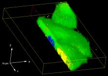
The Raman Microscope in the CCMR Bard Materials Facility now can make spatial maps of Raman spectra in three dimensions. The Volume Mapping feature for the WiRe 4.1 software from Renishaw provides a powerful new capability to survey through the depth of a specimen, making characterization of biological and mineralogical specimens much more effective. A recent publication describes 3D mapping of Raman signals in cells (http://pubs.rsc.org/en/content/articlehtml/2013/sc/c3sc51437d )
Volume Mapping uses the high-speed mapping mode StreamLineHR to acquire 3D maps over a period of minutes to hours, with the acquisition time depending on the size of the map and the strength of the Raman signal. StreamLineHR will also speed up two-dimensional maps. With StreamLineHR, Raman images can be acquired at a data rate of ~50 milliseconds per spectrum. This data rate is still 2 to 3 times slower than StreamLine Plus mode, which enables fast imaging by using a laser that is shaped into a line. However the line-mapping capability of StreamLine Plus cannot be used in 3D and has a spatial resolution limited by the CCD detector pixel size, so StreamLineHR does offer new capabilities relative to StreamLine Plus.
Curved or irregular surfaces can now be surveyed for topography in advance of Raman imaging so the focus is adjusted to keep the sample in focus during data acquisition. This automated focusing makes imaging large areas more user-friendly because the focus does not have to be adjusted manually.
Contact Dr. Kit Umbach of the Bard Materials Facility to learn more about Raman imaging.